
Dysplastic Nevi - Atypical Moles
The presence of dysplastic nevi can increase the risk of developing melanoma. However, not all dysplastic nevi progress to melanoma, and many remain benign throughout a person's life. Regular monitoring and evaluation by a dermatologist are essential for individuals with multiple dysplastic nevi, particularly those with a personal or family history of melanoma. Dermatologists may use dermoscopy, a technique involving a specialized magnifying device, to examine these moles more closely and identify any concerning changes. In some cases, a biopsy may be performed to rule out malignancy. Preventive measures, such as avoiding excessive sun exposure and using sunscreen, are crucial in reducing the risk of melanoma associated with dysplastic nevi.
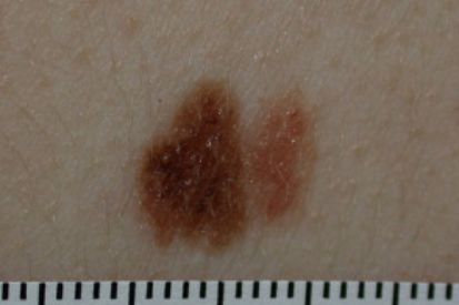
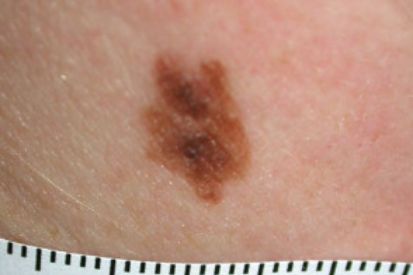
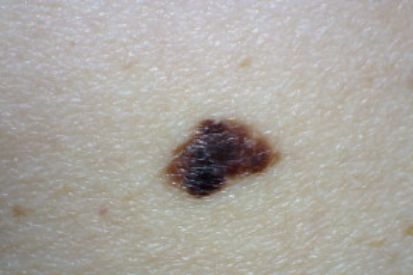
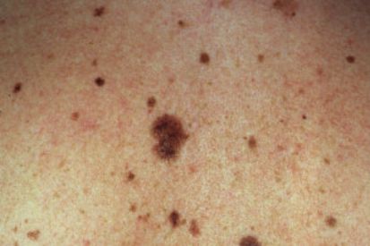
Examples of Dysplastic Nevi
What are the Symptoms of Dysplastic Nevi?
- Atypical moles are a size that exceeds that of common moles.
- These moles often exhibit a range of colors, including tan, brown, and red, creating an uneven or variegated appearance.
- The shape is irregular, featuring indistinct borders.
- They may appear either flat or slightly raised on the skin's surface, adding to their atypical nature.
- Dysplastic nevi are known for their potential to change over time. These changes can be alterations in size, color, or overall shape.
Causes of Dysplastic Nevi
- A family history of dysplastic nevi or melanoma indicates genetic predisposition.
- Exposure to UV radiation.
- Fair-skinned individuals with more moles are susceptible.
How to Prevent Dysplastic Nevi
- Sun Protection Measures: Wear protective clothing, including hats and long sleeves and use broad-spectrum sunscreen with a high SPF. Seek shade during peak sunlight hours between 10 a.m. and 4 p.m.
- Regular Skin Monitoring: Perform regular at-home self-exams of the skin and moles. Notify your dermatologist provider right away if you notice any unusual or evolving moles. It is also important to schedule an annual total body skin exam with one of our trusted providers.
- Awareness of Risk Factors: Individuals with a family history of dysplastic nevi or melanoma should be particularly vigilant, as they are at higher risk.
- Avoid Tanning Beds: Steer clear of tanning beds to reduce the risk of dysplastic nevi development.
FAQs for Dysplastic Nevi
Dysplastic nevi themselves are not cancerous; they are benign skin lesions. However, they are considered atypical moles due to their irregular appearance and potential to change over time. While most dysplastic nevi remain non-cancerous, their presence can indicate an increased risk of developing melanoma, a serious form of skin cancer. This risk is particularly heightened in individuals with multiple dysplastic nevi or a family history of melanoma.
Regular monitoring and dermatological evaluations are essential for individuals with dysplastic nevi to detect any changes early. Dermatologists may use dermoscopy to closely examine these moles for any signs of malignancy. If any suspicious changes are observed, a biopsy may be performed to determine if the mole has become cancerous. Preventative measures, such as minimizing sun exposure and consistently using sunscreen, are also important in managing the risk associated with dysplastic nevi.
Dysplastic nevi are diagnosed through a combination of visual examination and dermoscopic analysis by a dermatologist. During a skin examination, the dermatologist looks for moles that exhibit asymmetry, irregular borders, multiple colors, and larger size compared to common moles. Dermoscopy, a technique using a specialized magnifying device, allows the dermatologist to see structures and patterns not visible to the naked eye, aiding in the identification of atypical features. If a mole appears particularly suspicious, a biopsy may be performed, where a small sample of the mole is removed and examined under a microscope to check for cancerous cells.
Monitoring dysplastic nevi involves regular skin checks, both self-examinations by the patient and periodic professional evaluations by a dermatologist. Individuals with multiple dysplastic nevi or a family history of melanoma should have more frequent check-ups. Digital dermoscopy and photography can be used to track changes in moles over time, providing a visual record that helps in detecting any new developments or alterations. This proactive monitoring is crucial for early detection of melanoma, allowing for timely intervention and treatment if needed. Preventative measures, such as protecting the skin from excessive sun exposure and using sunscreen, are also recommended to reduce risks associated with dysplastic nevi.
Dysplastic nevi have the potential to turn into melanoma, although this transformation is not very common. These atypical moles are considered a marker for an increased risk of melanoma, especially in individuals with a large number of dysplastic nevi or a family history of melanoma. The irregular shape, color variation, and asymmetry of dysplastic nevi can sometimes make it difficult to distinguish them from early melanomas, necessitating careful and regular monitoring.
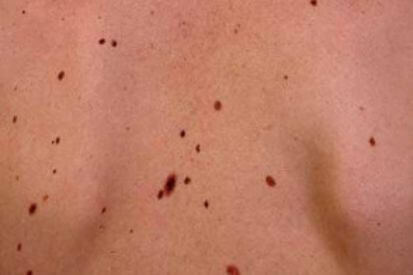
Yes, dysplastic nevi can develop anywhere on the body, although they are more commonly found on sun-exposed areas. These areas include the back, chest, and limbs, where the skin is more frequently exposed to ultraviolet (UV) radiation from the sun. Despite their common occurrence in these sun-exposed regions, dysplastic nevi can also appear in less exposed areas such as the scalp, buttocks, and even the soles of the feet or palms of the hands.
A total body skin exam by a dermatologist is crucial for the early detection of skin cancers, including melanoma, basal cell carcinoma, and squamous cell carcinoma. Dermatologists are trained to identify suspicious moles and skin lesions that might be overlooked during self-examinations. They use specialized tools like dermoscopy to examine the skin in detail, which allows them to detect abnormalities that are not visible to the naked eye. Early detection is vital because it significantly increases the chances of successful treatment, particularly for aggressive cancers like melanoma, which can spread rapidly if not caught early.
Moreover, a total body skin exam can help in monitoring individuals with a high number of moles or those with dysplastic nevi, who are at an increased risk of developing skin cancer. During these exams, dermatologists document the appearance of moles and track any changes over time, using techniques such as digital dermoscopy and photography. This systematic approach ensures that any new or evolving lesions are promptly identified and evaluated. Regular skin exams also provide an opportunity for dermatologists to educate patients about proper skin care and preventive measures, such as the use of sunscreen and the avoidance of excessive sun exposure, further reducing the risk of skin cancer.
From Our QualDerm Family of Brands: Healthy Skin with Total Body Skin Exams
How to Treat Dysplastic Nevi
- Surveillance and Monitoring: One of our dermatologists will closely monitor dysplastic nevi due to their potential to transform into melanoma. Regular at-home self-exams are key – make sure to notify your dermatologist of any unusual or evolving moles. It is also important to schedule regular Total Body Skin Exams at one of our locations.
- Surgical Removal: If dysplastic nevi exhibits concerning features or irregularities during our providers’ evaluations, your dermatologist may recommend an excisional biopsy or complete removal.
- Reducing Malignancy Risk: Excisional biopsy aims to reduce the risk of potential malignancy by removing atypical moles.
- Regular Follow-ups: Individuals with a history of dysplastic nevi or a family history of melanoma should maintain regular visits with one of our skincare experts.
- Optimal Skin Health: While not all dysplastic nevi require treatment, their careful monitoring is crucial for optimal skin health and cancer prevention.
Related Blog Posts

- Skin Cancer
- General Dermatology
- Chronic Skin Conditions
Learn more about the most common types of skin lesions we see at Cumberland Skin and how our dermatologists remove them.
Read More
- General Dermatology
- Skin Exams
Preparing for your first dermatology appointment is important because it ensures everything goes as smoothly as possible and that your doctor is up-to-date on the status of your overall health and wellbeing. Here are our expert tips.
Read More
- Skin Cancer
- Skin Exams
Discover the ABCDEs of melanoma. Familiarize yourself with the five key indicators to aid in early detection and prompt medical attention for any suspicious moles or skin lesions.
Read MoreFeatured Products for Sun Protection
Check your local office for current stock!
Check your local office for current stock!